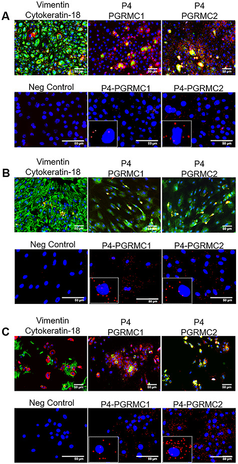
Figure 4.

Proximity ligation assay shows P4 binding to both PGRMCs in all fetal membrane cell types. Top left panel: intermediate filament markers vimentin (mesenchymal marker; green) and cytokeratin-18 (epithelial marker; red) confirm primary cell type. Middle and left panel: immunocytochemistry controls validating P4 (red), PGRMC1 (green), and PGRMC2 (green) antibodies. Bottom left panel: positive and negative probes only without primary antibodies to act as a negative (Neg) control. (A) P4 bound directly to PGRMC1 and PGRMC2 in AECs (as seen by red dots); however, P4 binding was more significant to PGRMC2 (N = 3). (B) P4 bound directly to both PGRMC1 and PGRMC2, as in AMCs (N = 3). (C) Chorion cells exhibited the most P4 receptor interactions of all cells types, but did not significantly prefer PGRMC2 to PGRMC1. Scale bar is 50 μm (N = 3).
