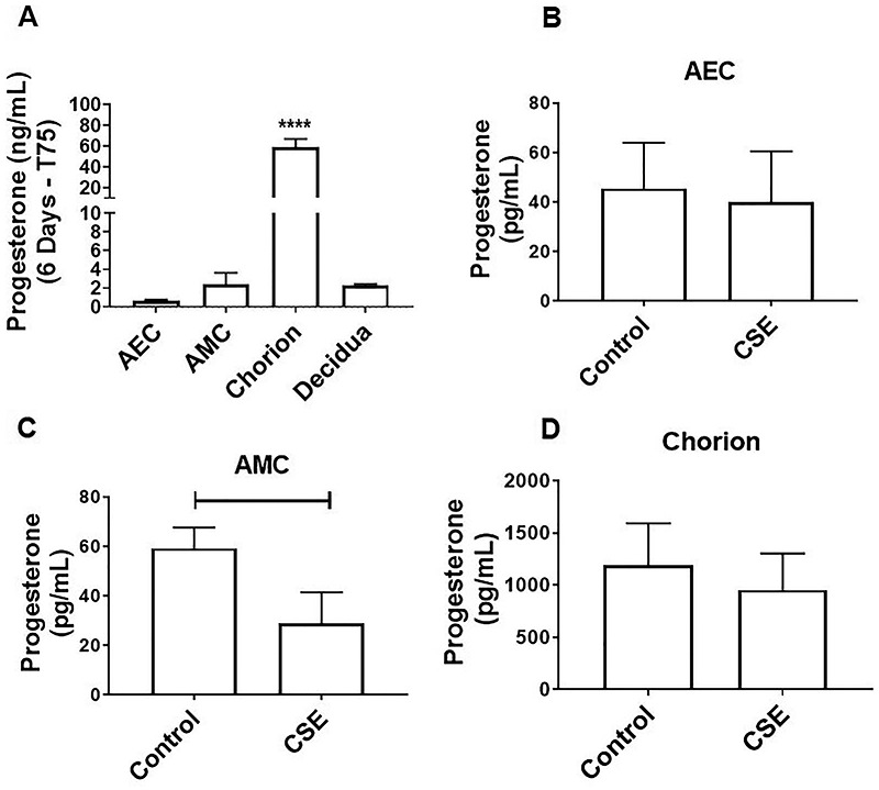
Figure 5.

Oxidative stress inhibits local P4 release in AMCs. (A) Although all fetal membrane cells can produce progesterone, chorion cells produce higher levels compared with amnion epithelial and mesenchymal cells (AEC: 0.6 ± 0.1 ng/mL, AMC: 2.5 ± 1.2 ng/mL, chorion: 58.8 ± 8.1 ng/mL). In addition, full fetal membrane explants secrete significant levels of P4, which is likely produced by the chorion layer (P < 0.0001) (chorion: 58.8 ± 8.1 ng/mL, decidua: 2.3 ± 0.1 ng/mL) (N = 3; mean ± SEM). To compare cell types under normal cell culture conditions, we extrapolated and normalized P4 production for 6 days within a T75 flask. (B and D) CSE-induced oxidative stress did not affect P4 production in AECs (B), CSE decreased P4 production in AMCs (C) (P = 0.039; control: 59.2 ± 8.43 pg/mL, CSE: 28.95 ± 12.5 pg/mL) and chorion cells (D) (control: 1192 ± 401.6 pg/mL, CSE: 953.9 ± 350 pg/mL) (N = 3; mean ± SEM).
