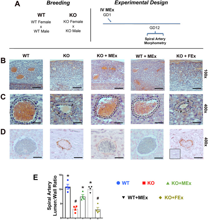
Figure 2.

Spiral artery morphology is altered by antenatal MEx treatment. (A) Experimental design. (B, C) Representative H&E images of spiral arteries at (B) low magnification (×100) and (C) high magnification (×400). Dotted lines denote area of analysis for lumen:wall ratios. Stars denote thickened blood vessel walls. (D) Representative images of smooth muscle actin immunohistochemistry. Red stain: smooth muscle actin; blue stain: hematoxylin nuclear stain. Inset black box: second only negative control. (E) Graphical analysis representative of four independent experiments, total n = 12–16 placentas for each condition. Scale bars ×100: 50 μm, ×400: 15 μm. Statistical significance (P < 0.05) as determined by one-way analysis of variance is denoted by different symbols (*, #).
