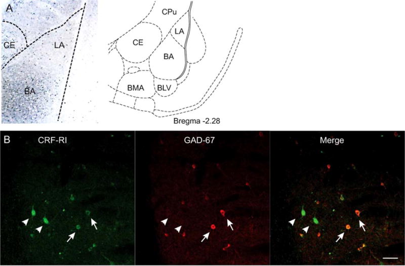
FIGURE 1.

Localization of CRF1 receptors on GABAergic interneurons. (A) Left: distribution of CRF1 receptors in the amygdala. LA: lateral nucleus, BA: basal nucleus, CE: central nucleus. Right, samples were taken from the LA and BA. CPu: caudate putamen, BLV: ventral basolateral nucleus, BMA: basomedial nucleus. Adapted from Paxinos and Watson (1998). (B) Colocalization of CRF1 receptors (green) and GAD67 (red). Merging of the red and green channels indicates several somata expressing both GAD67 and CRF1 receptors (arrows). Several neurons expressing CRF1 receptors (arrowheads) do not express GAD67. Scale bar: 50 μm
