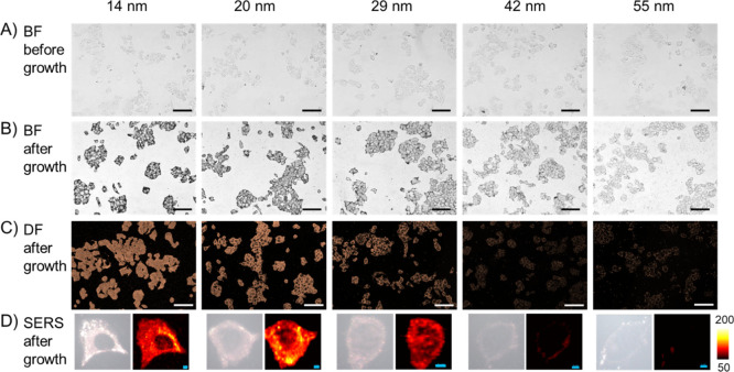
Figure 2.
Penetration of nanoprobes into methanol-fixed MCF-7 cells. (A) Bright-field images of cells incubated with nanoprobes of different sizes. (B) Bright-field images and (C) dark-field images of the cells after adding the Au growth solution. (D) Typical SERS scanning images of the cells incubated with nanoprobes of different sizes and after growth. Scalebar in BF/DF images: 100 μm; scalebar in SERS images: 4 μm.

