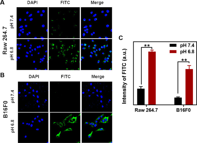
Figure 3.
Uptake of Cu-LDH@PAMA/DMMA by B16F0 and Raw 264.7 cells. CLSM images of Raw 264.7 cells (A) and B16F0 cancer cells (B) and their mean fluorescence intensity (C) via flow cytometric analysis after the cells were treated with Cu-LDH@PAMA/DMMA in media with pH 7.4 and 6.8 at 37 °C for 4 h. **p < 0.01.

