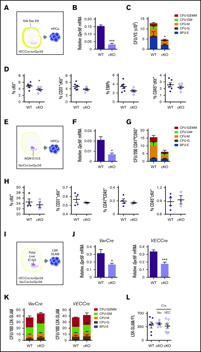
Figure 1.
Gpr56 deficiency effects early hematopoietic development in mouse embryos. Experimental setup for E9 YS (21-25 somite pairs) (A), E10.5 AGM (34-37 somite pairs) (E), and E13.5 FL HPC analyses (I). (B) Relative expression of Gpr56 in WT and VECCre:loxGpr56 cKO E9 YS cells normalized to β-actin by RT-PCR analysis (n = 3). (C) Number of CFU per WT and cKO E9 YS (n = 3). (D) Percentages of cKit+, CD31+cKit+, CD41+cKit+CD16/32+ (EMP), and CD45+ckit+ cells in WT and cKO E9 YS (n = 6). Flow cytometric gating strategy shown in supplemental Figure 1. (F) Relative expression of Gpr56 in WT and VavCre:loxGpr56 cKO E10.5 AGM cells normalized to β-actin by RT-PCR analysis (n = 3). (G) Number of CFU per WT and cKO E10.5 AGM (n = 3). (H) Percentages of cKit+, CD31+cKit+, CD41loCD45+, and CD45+ckit+ cells in WT and cKO E10.5 AGMs (n = 6). (J) Relative expression of Gpr56 in WT and VavCre: and VECCre:loxGpr56 cKO LSK SLAM sorted E13.5 FL cells normalized to β-actin by RT-PCR analysis (n = 5). *P ≤ .05. (K) Number of CFU per WT and Vav and VECCre cKO E13.5 FL LSK SLAM cells (n = 4). (L) Number of LSK SLAM cells per WT and Vav and VECCre cKO E13.5 FL (n = 6). Distinct colony types are indicated. Mean ± standard error of the mean (SEM) is shown.

