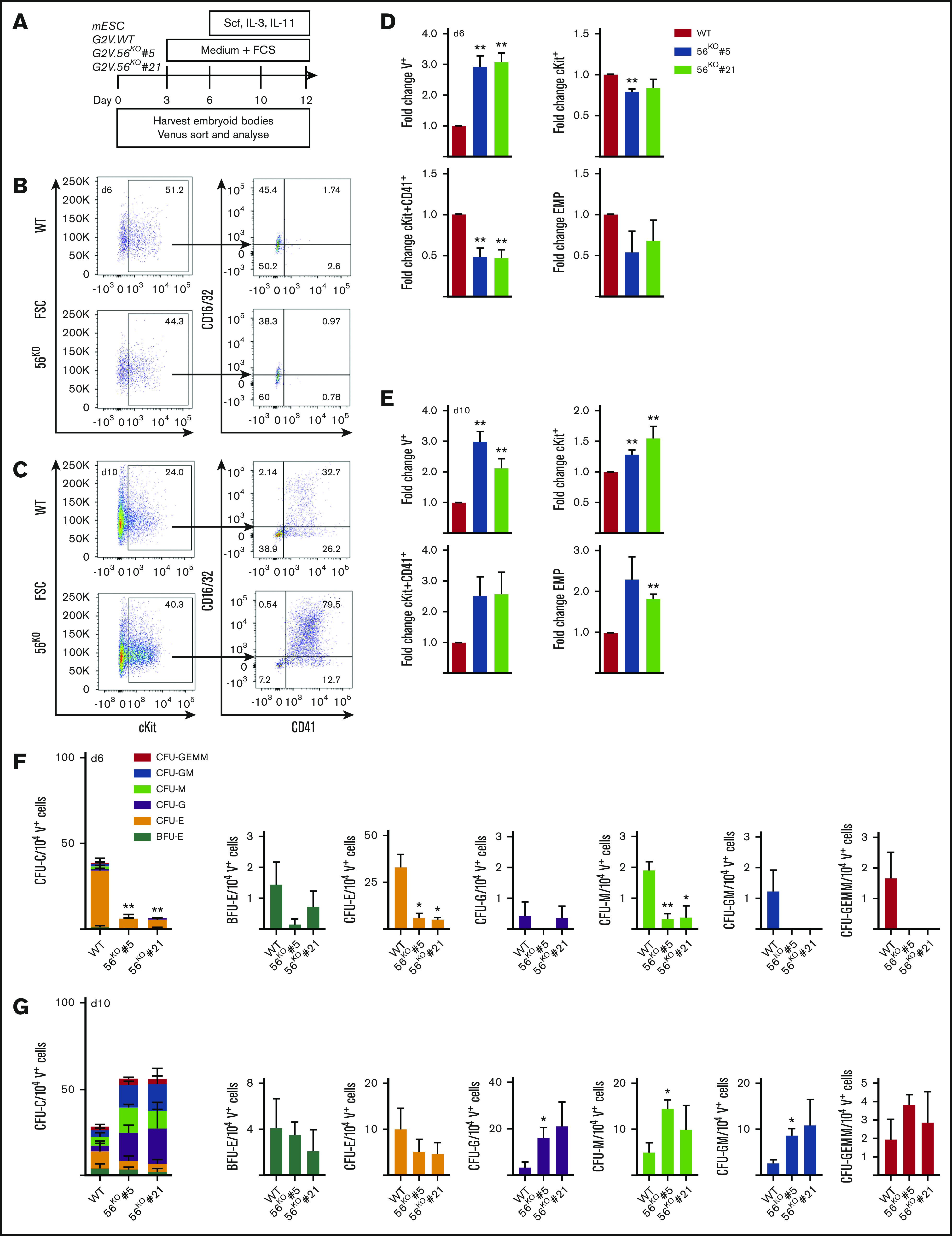
Figure 3.

Gpr56 affects hematopoietic output in vitro. (A) ESC differentiation culture methodology. Mouse Gata2Venus (mG2V) ESC (WT and 56KO) were differentiated in hematopoietic factor-containing medium for several days and unsorted/sorted V+ and V− cells were examined for Gpr56 mRNA (RT-PCR) and Gpr56 protein (WB, western blot) expression. (B-C) Representative flow cytometric plots of day 6 (B) and day 10 (C) G2V.WT and G2V.56KO differentiation cultures showing percentages of CD16/32 and CD41 cells in Venus+cKit+ gate. CD41+cKit+ CD16/32+ = EMP (erythromyeloid progenitors). (D-E) Fold change in the percentages (mean ± SEM) of Venus+, cKit+, cKit+CD41+, and EMPs in day 6 (D) and day 10 (E) G2V.WT and G2V.56KO (clones #5 and #21) differentiation cultures. n = 3. (F-G) Hematopoietic potential of G2V.WT and G2V.56 KO (clones #5 and #21) HPCs was determined at day 6 (F) and day 10 (G) of differentiation by CFU-C assay. CFU-C per 10 000 V+ plated cells is shown. Distinct colony types are indicated by color. n = 3; mean ± SEM.
