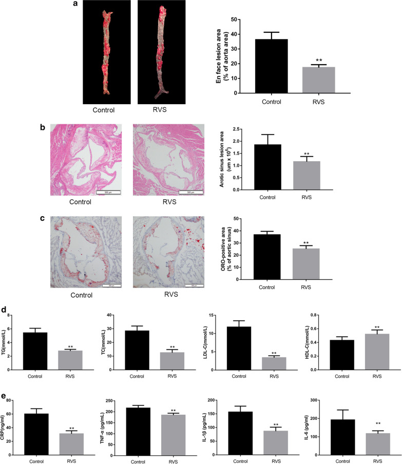
Fig. 1.
The effects of RVS on the progress of atherosclerotic plaques, blood lipid parameters and the content of inflammatory cytokines in vivo. a The lesion area of entire aorta was measured via ORO staining. b The size of atheroma plaques in cross-sections of aortic sinus was detected by HE staining. c The representative lipid deposition in cryosections of aortic sinus were stained with ORO. d The serum level of TG, TC, LDL-C and HDL-C was examined by the corresponding kits. e The level of CRP, TNF-α, IL-1β and IL-6 was assayed by ELISA kits. Data were expressed as the mean ± SD, n = 6. *p < 0.05, **p < 0.01 vs. the Control group

