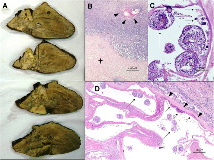
Fig. 2.
Patient No. 14. a Gross view of the resected right lobe of the liver shows heterogeneous solid mass lesion that contains externally budding vesicles and calcified foci with a maximum diameter of a single vesicle around 10 mm. b, c, d Histopathological characteristics of Echinococcus multilocularis lesion in the liver. Normal appearance of the liver is distorted. Severe inflammation is present around the necrotic liver tissue (asterisk in b). This inflammatory infiltrate consists of lymphocytes and histiocytes. In some areas fragment of cuticular membrane is observed within the necrotic area (arrowheads in b). This membrane displays the laminated layer as a tender band-like structure (arrowheads in d) with a germinal layer (short arrows in c and d). Note the invaginated protoscoleces found within the vesicles (long arrows in c and d)

