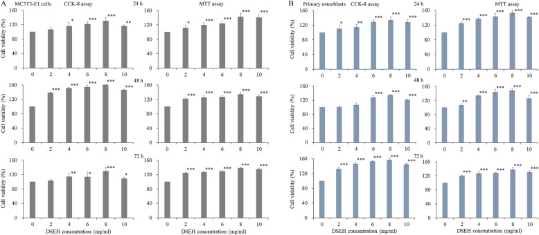
Fig. 1.
Effects of DSEH on the cell proliferation of MC3T3-E1 cells and primary osteoblasts. a MC3T3-E1 cells. b Primary osteoblasts. Cell viabilities were detected by CCK-8 and MTT assays under the treatment of DSEH at progressively increasing concentrations of 0 mg/ml, 2 mg/ml, 4 mg/ml, 6 mg/ml, 8 mg/ml and 10 mg/ml) at different time points (24 h, 48 h, 72 h). Cell viabilities of the DSEH treatment groups under different concentrations were estimated by normalizing to that of the untreated group (0 mg/ml). Data were presented as the mean with standard deviation for technical triplicate in an experiment representative of several independent ones (n = 4). p < 0.05, p < 0.01 and p < 0.001 represented the differences in cell viabilities under DSEH treatment in a student t-test

