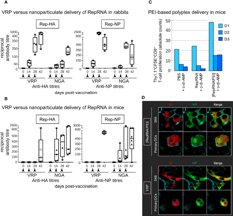
Figure 2.
Virus replicon particle (VRP) and NGA deliver RepRNA for translation in vivo. (A, B) Mice and rabbits were injected subcutaneously at 0, 14, and 28 days. Rep-HA and Rep-NP were delivered in NGA or VRP; all vaccines were adjuvanted with c-di-AMP adjuvant. Serum samples were assessed for anti-HA and anti-NP antibodies by ELISA and titers estimated at the times shown. (C) Adoptive transfer studies in mice have been performed in order to investigate the potential of the polyethylenimine (PEI) formulations to stimulate cellular immune responses. In brief, 24 h prior vaccination of wild type mice, CFSE labeled Thy1.1 CD8+ T cells of antigen-specific TCR transgenic mice were injected intravenously. 7 days later, cervical LNs of transplanted mice were collected and the proliferative capacity of the CFSE labeled Thy1.1 CD8+ T cells was analyzed by flow cytometry. Cell number as well as the number of cell divisions correlate with both strength of the stimulated cellular response and vaccine delivery efficacy. The number of division cycles (D1-D3) of proliferating OT-I CD8+ T cells were determined by flow cytometry (FCM) using the FlowJo software. For each group, tissues from five mice were pooled to provide enough cells. (D) Translation of delivered Rep-NP in porcine SK-6 cells and primary dendritic cells (DCs). After incubation for 48 h at 37°C with Rep-NP complexed to PEI or packaged into VRP, the cells were washed, fixed (4% PFA), permeabilised (saponin) and labeled with antibodies against influenza NP (green) and CSFV E2 (red); cell surfaces were stained with WGA (blue). All pictures were generated with IMARIS 7.7 with threshold subtraction and gamma correction set as in the “RepRNA alone” control.

