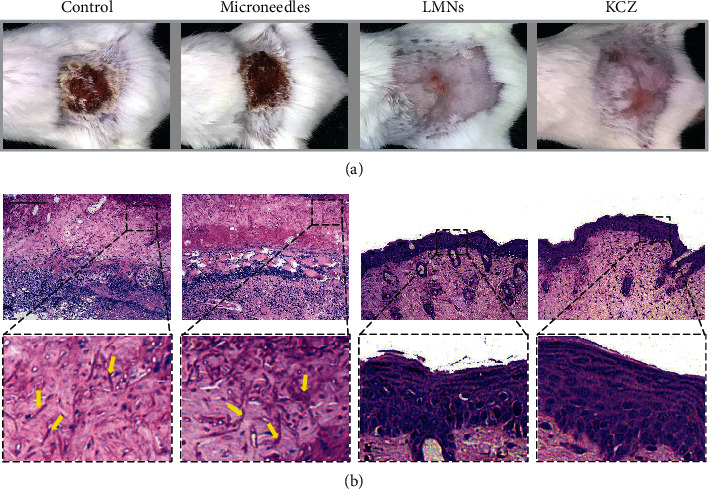
Figure 4.

The in vivo antifungal capability of the LMNs. (a) Representative photos of the fungi-infected skin treated with PBS, microneedles, LMNs, and KCZ. (b) PAS staining of fungi-infected tissue after 14 d. Yellow arrows indicated the pseudohyphae of C. albicans. Scale bar in (b) was 100 μm.
