Figure 5.

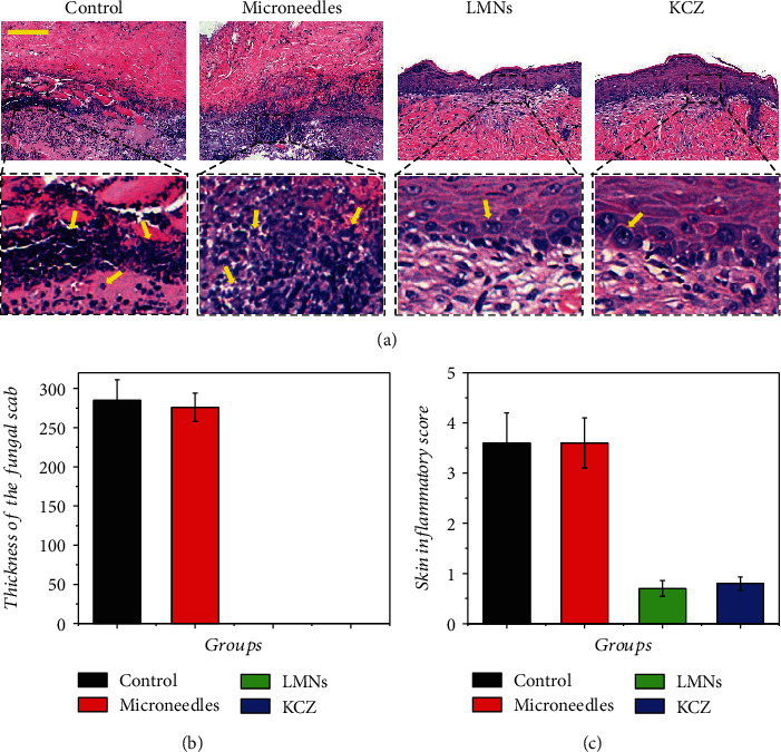
(a) HE staining of fungus-infected tissue after 14 d. (b) Quantification of thickness of the fungal scab. (c) Quantification of the skin inflammatory score. Yellow arrows indicated the inflammatory cells. Scale bar in (a) was 100 μm.

(a) HE staining of fungus-infected tissue after 14 d. (b) Quantification of thickness of the fungal scab. (c) Quantification of the skin inflammatory score. Yellow arrows indicated the inflammatory cells. Scale bar in (a) was 100 μm.