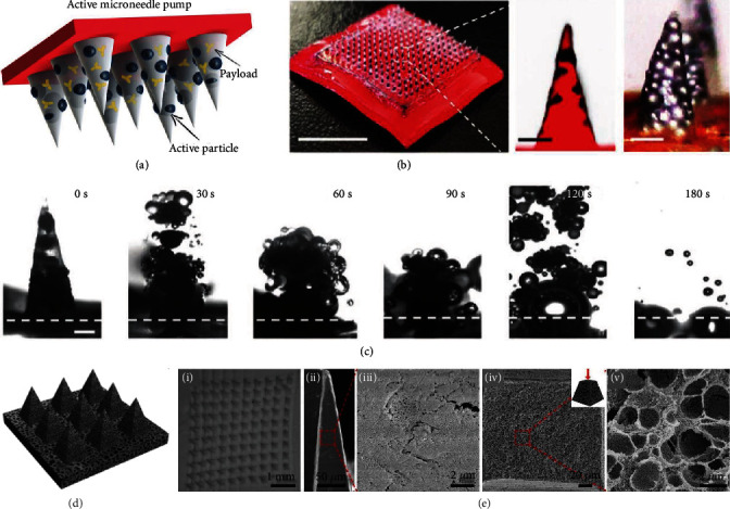
Figure 5.

Explosive and porous microneedles. (a) Schematic illustration of the composition of active microneedles. (b) Digital photo of an active microneedle patch and optical/fluorescence microscopy images of an active microneedle tip. The scale bars are 300 μm and 25 μm, respectively. (c) Real-time images of the explosion of a single active microneedle tip in PBS solution. The scale bar is 200 μm. Reproduced with permission from Ref. [58], copyright 2020, Wiley-VCH. (d) Schematic illustration of porous polymer microneedles. (e) SEM images of a porous microneedle array (i), the surfaces (ii, iii), and the cross-sections (iv, v) of a porous microneedle tip. Reproduced with permission from Ref. [59], copyright 2020, Royal Society of Chemistry.
