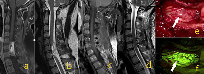
Figure 1.
Schwannoma at the C6-7 level in preoperative contrast-enhanced T1 and T2 sequences on images (A, B). On postoperative (C, D) images, the mass was totally removed. In intraoperative microscope image (E) white arrow shows the mass, while image (F) shows tumor homogeneous staining pattern with activated yellow filter.

