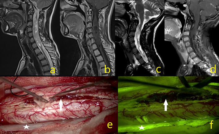
Figure 4.
A 23-year-old male patient operated for oligodendroglioma WHO grade 2, but where subtotal excision could not be achieved due to intraoperative IONM signal loss. (A–D) Tumor MR sequence images. (E) In the intraoperative microscope image, the asterisk is the dura and the white arrow indicates the mass. (F) Under the yellow filter, the asterisk shows the dura staining pattern, and the white arrow a moderate heterogeneous stained mass image.

