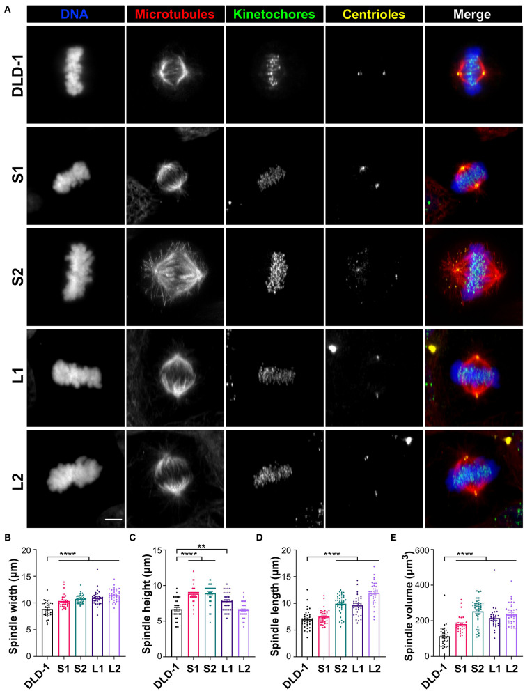
Figure 2.
Tetraploid DLD-1 clones have distinct mitotic spindle geometries. (A) Examples of mitotic spindles from parental DLD-1 cells and cells from 4N DLD-1 clones S1, S2, L1, and L2 (top-to-bottom) in metaphase. Scale bar, 5 μm. Measurements of mitotic spindle width (B), height (C), length (D), and volume (E) reported as mean ± SEM with individual data points from three independent experiments in which a total of 30–40 cells (n = 35, 30, 40, 30, and 35, respectively). **p < 0.01, ****p < 0.0001, when compared to the parental DLD-1 cells by Student's t-test.

