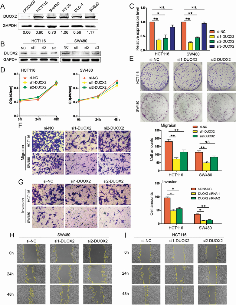
Figure 2.
Knockdown of DUOX2 decreases the migration and invasion of CRC cells in vitro. (A) DUOX2 protein expression levels in one normal intestinal epithelium cell line and five CRC cell lines. (B and C) Western blotting and qRT-PCR were performed to detect the DUOX2 expression levels in DUOX2 knockdown (si1-DUOX2, si2-DUOX2 and si3-DUOX2) and negative control (si-NC). *P < 0.05, **P < 0.01, N.S.P > 0.05. (D) MTS assay. DUOX2 knockdown did not attenuate the cell viability of HCT116 and SW480 cells. Each point indicates the mean of spectrometric absorbance OD492 ± SD of three independent experiments. (E) Colony assays. DUOX2 knockdown did not attenuate colony formation in HCT116 and SW480 cells. (F) Transwell migration assays. (G) Transwell invasion assays. The error bars represent the standard deviation. *P < 0.05, **P < 0.01, N.S.P > 0.05. (H and I) Wound healing assay. The migration rate was derived from the ratio of the difference in wound area at different times to the initial wound area (200×). The tests were performed on SW480 and HCT116 cells. *P < 0.05, **P < 0.01.

