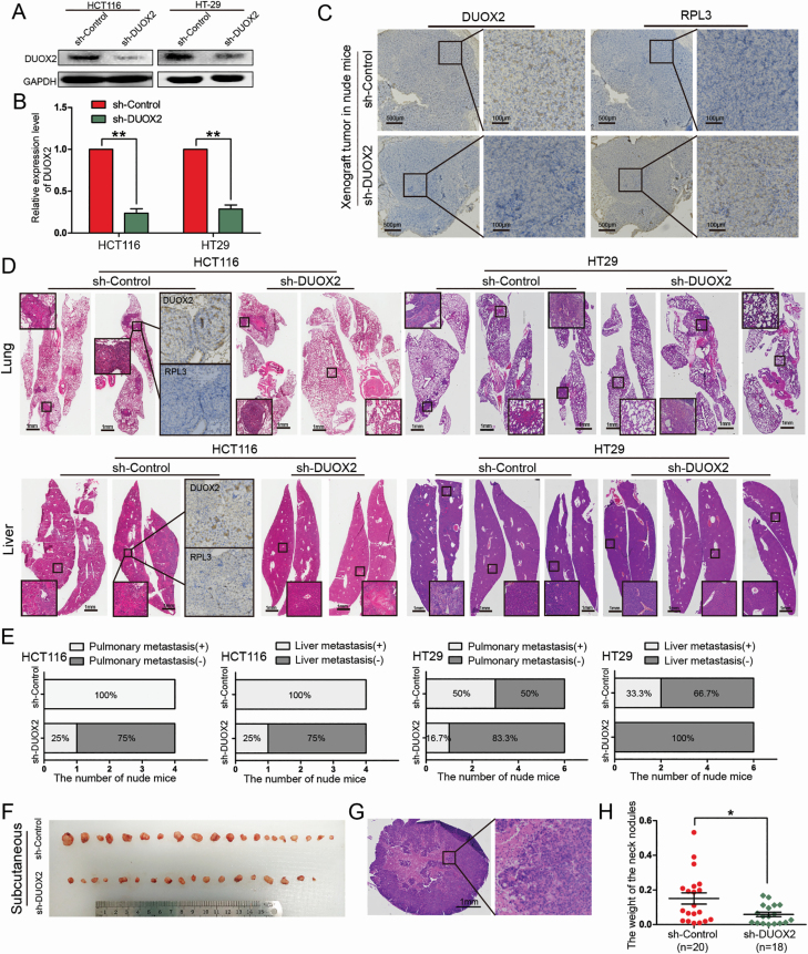
Figure 5.
Knockdown of DUOX2 inhibits the ability of tumor invasion and metastasis in vivo. (A and B) Western blotting and qRT-PCR were performed to detect the DUOX2 expression levels in DUOX2 knockdown (sh-DUOX2) and negative control (sh-Control). **P < 0.01. (C) IHC detection of DUOX2 and RPL3 proteins in xenograft tumor of nude mice, which were injected sh-DUOX2 or sh-Control HCT116 cells. (D) All the lung and liver tissues of different groups were extracted and stained with HE staining. IHC test was preformed to detect the expression of DUOX2 and RPL3 proteins in lung metastases and liver metastases tissues. (E) Statistical analysis of lung and liver metastasis in different groups. (F) After injection of HT29 cells into the tail vein, significant subcutaneous nodules were found in the neck of nude mice. (H) The weight of subcutaneous metastatic tumors in sh-DUOX2 group was significantly lower than those in sh-Control group (P = 0.014). (G) These nodules were stained with HE. *P < 0.05), **P < 0.01.

