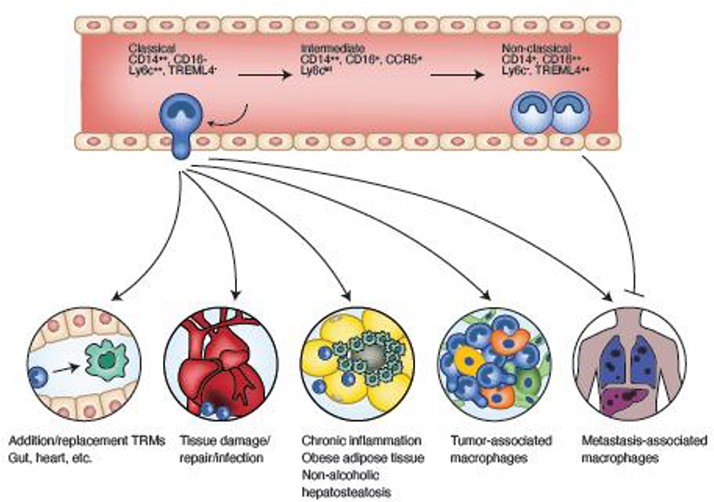
Figure 1. Monocyte differentiation in blood and tissues.
Classical monocytes differentiate into non-classical monocytes through an intermediary stage. Markers are shown for human (top) and mouse (bottom). Classical monocytes extravasate into tissues to replace resident macrophages, participate in wound responses, contribute to pathogenic tissue inflammation, and promote tumor progression and metastasis. Non-classical monocytes are retained in vessels where they perform surveillance and monitor pathogenic insults. They can also extravasate at sites of metastasis and inhibit tumor cell establishment through a NK cell mediate mechanism.

