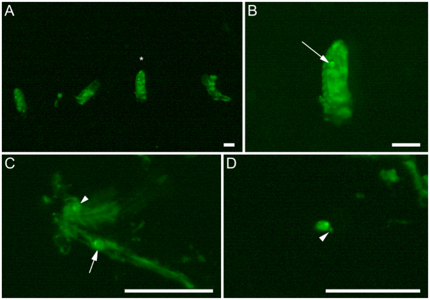
Figure 3: The dissection process.

(A–D) Embryos that express six4-eGFP::moesin (green) at sequential stages in the dissection protocol. (A) Four devitellinized embryos that have been transferred to the poly-lysine-coated dissection region of the dish. Note how the embryos are aligned in a neat row to facilitate successive downward dissections in the dish. The small piece of tissue between Embryos 1 and 2 is part of the gut from Embryo 2, extruded during hand devitellinization. (B) Higher magnification view of embryo from (A), indicated by the asterisk. (C) The remaining carcass of an embryo that has been filleted down the midline. The arrowhead indicates an occluded gonad, still heavily embedded in embryonic tissues. (D) A completely dissected gonad. Note only minimal extraneous tissue (arrowhead) remains near the gonad. Arrows point to male gonads. Scale bars are 0.25 mm.
