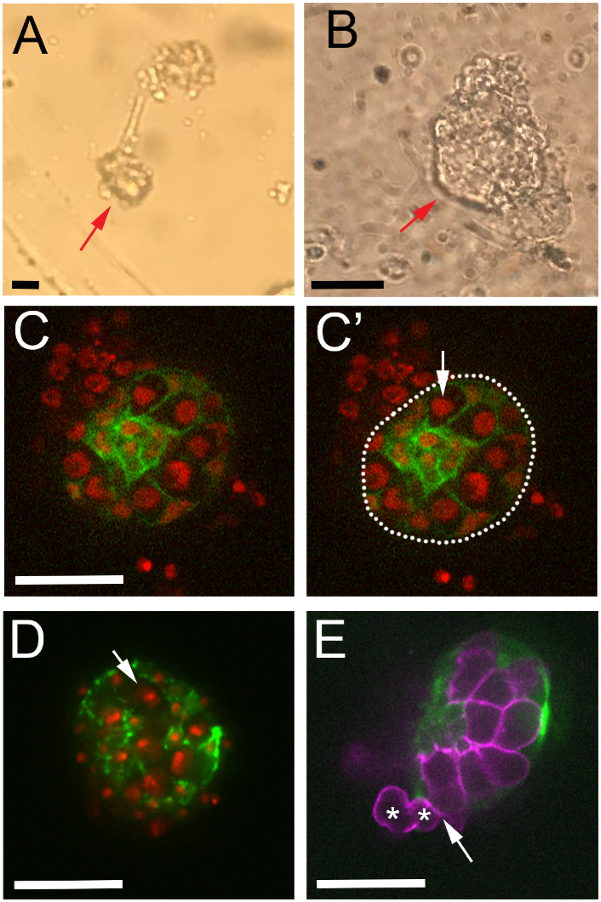
Figure 4: Locating healthy gonads during live-imaging.

(A) Low and (B) high magnification brightfield views of two dissected gonads adhered to the coverslip. (C–C’) One movie frame from ex vivo imaging of niche compaction in a healthy gonad. Somatic gonadal cells express six4-eGFP::moesin (green), and all cells express His2Av-mRFP (red). (C’) Gonad boundary marked with a white dotted line. His2Av-mRFP visible outside of the gonad boundary is likely fat body that isstill attached to the dissected gonad. Arrow points to a germ cell nucleus with uniform His2Av-mRFP signal, indicating the gonad is healthy. (D–E) Representative negative outcomes of the ex vivo dissection protocol. (D) A frame from an imaging session in which the gonad has dehydrated because of media evaporation. Note pyknotic nuclei as His2Av-mRFP condenses (arrow), and discontinuous spots of six4-eGFP::moesin along the gonad boundary. (E) Ex vivo imaging framein which extracellular matrix has been compromised during dissection. Note that some germ cells (asterisks) are exiting the gonad boundary (arrow). Germ cells are labeled with nos-lifeact::tdtomato (magenta), and somatic gonadal cells with six4-eGFP::moesin (green). Scale bars show 20 μm.
