
Figure 4.
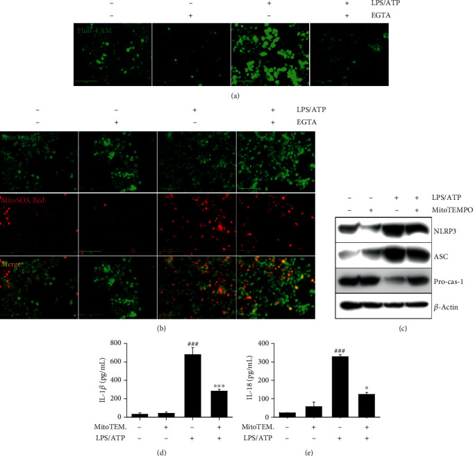
LPS/ATP-induced intracellular Ca2+ accumulation activates the NLRP3 inflammasome through mtROS production. BV2 microglia cells were pretreated with 2 mM EGTA for 2 h prior to stimulation with LPS/ATP for 12 h. The cells were stained with (a) 1 μM Fluo-4 AM and (b) 0.5 μM MitoTracker Green and 2 μM MitoSOX Red. Cell images were captured by CELENA S Digital Imaging System. (c) The cells were pretreated with 50 μM MitoTEMPO for 2 h prior to stimulation with LPS/ATP. Total protein was isolated at 12 h, and western blotting was performed. Cell culture supernatants were collected at 48 h, and ELISA was performed to quantify the levels of (d) IL-1β and (e) IL-18. ∗∗∗p < 0.001 and ∗p < 0.05 vs. untreated cells; ###p < 0.001 vs. LPS/ATP-treated cells.