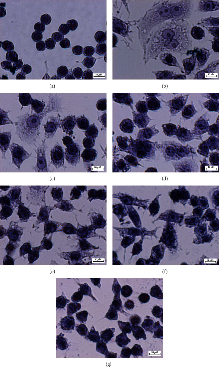
Figure 3.

Effects of GIE on the morphology of LPS plus IFN-γ-induced RAW264.7 cells. Cells were stained with hematoxylin staining: (a) uninduced RAW264.7 cells, (b) untreated LPS plus IFN-γ-induced cells, (c) cells were pretreated with DEX at 1 μM, (d), (e), (f), and (g) cells were pretreated with GIE at concentrations range of 50, 100, 200, and 300 μg/mL, respectively (original magnification at ×600, scale bar; 10 μm).
