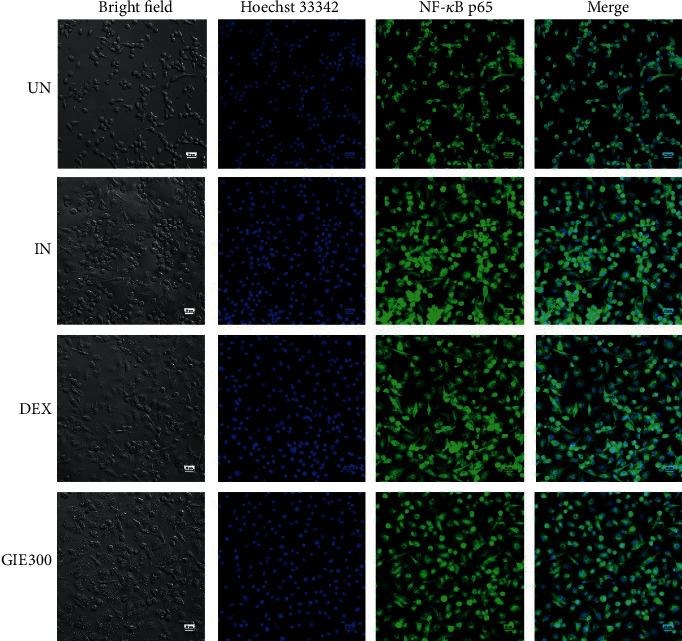
Figure 4.

Effects of GIE on the nuclear translocation of NF-κB p65 in LPS plus IFN-γ-induced RAW264.7 cells at 24 h. Cells were pretreated with GIE or DEX for 3 h and then coincubated with LPS plus IFN-γ for 24 h. The nuclear translocation of NF-κB p65 was detected using an immunofluorescence assay and visualized under confocal microscopy. The figure represents the cell morphology (bright field), the nuclear translocation of NF-κB p65 (green fluorescence), nucleus (blue fluorescence), and costaining (overlay green and blue fluorescence). Scale bar, 20 μm. UN: uninduced cells; IN: untreated LPS plus IFN-γ-induced cells; DEX: cells were pretreated with DEX at 1 μM; GIE300: cells were pretreated with GIE 300 μg/mL.
