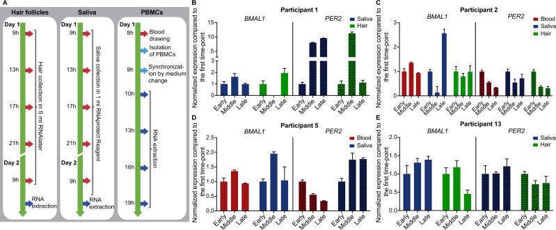
Figure 1.
Experimental methodology and sampling schemes for circadian rhythmicity for different human peripheral tissues. (A) RNA for time-course RT-qPCR was extracted (dark blue arrows) from human hair follicles, human saliva and human PBMCs. Red arrows denote sampling time points. (B–E) Three time-point comparison of BMAL1 and PER2 expression for participants 1, 2, 5 and 13 (aged 30.60±7.02 years; mean, SD). Expression data are normalised to the first time-point (early). For hair and saliva data early, middle and late time-points represent 9h, 17h and 21h, respectively. For PBMCs data early, middle and late time-points represent 10h, 16h and 19h, respectively. Depicted are mean±SEM, error bars represent technical replicates. PBMCs, peripheral blood mononuclear cells.

