Figure 2.
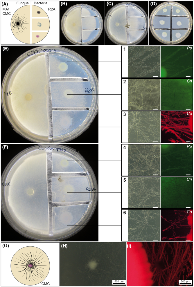
Validation of the ability of bacteria to colonize the mycelial network of the fungus C. cinerea. Dispersal of fluorescently labeled Cupriavidus oxalaticus (Co), Cupriavidus necator (Cn) and Pseudomonas putida (Pp) was tested in two-compartment Petri dishes with malt agar (MA) and carboxymethyl cellulose (CMC) (fungal growth), and R2A (bacterial growth) media or by co-inoculation in CMC. (A) Schematic representation of the experimental design in which the fungus and bacteria were inoculated in physically separated compartments. To test bacterial dispersal, agar pieces containing the bacterial inoculum were physically separated by cutting out agar slices forming a gap that must be connected by fungal hyphae. Macroscopic images demonstrating the colonization by C. cinerea grown on MA (B) or CMC (C) of the R2A media without bacteria. (D) Control with bacteria only grown on R2A. Macroscopic images demonstrating the colonization by C. cinerea grown on MA (E) or CMC (F) of R2A media pre-inoculated with bacteria. Bacterial dispersal was visually assessed by stereoscopic observations (right-hand panels). The colocalization of the fluorescence with the hyphae (bright field) indicates the colonization of the fungal mycelial network by the fluorescently labeled bacteria. The pictures correspond to close-up images taken from the R2A medium. The scale bar in the close-up images (in white) corresponds to 50 µm. (G) Schematic representation of the experimental design in which the fungus and bacterium were co-inoculated directly in the same medium. (H and I) Images showing the colonization of the fungal mycelium when C. cinerea was co-inoculated in CMC together with C. oxalaticus. The white spot observed in the bright-field image corresponds to a fungal primordium, which was not colonized by the bacteria as seen in the fluorescent image.

