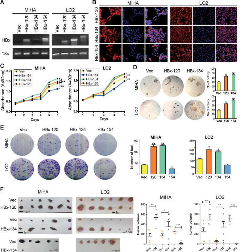
Fig. 2. Ct-HBx promotes hepatocarcinogenesis.
A RT-PCR was applied for validation of overexpression of HBx at the genomic level, 18 s served as an internal reference. B Immunofluorescence staining was applied for validation of overexpression of HBx at the protein level and the subcellular location of HBx was also indicated. C Ct-HBx promotes cell viability as indicated by XTT value in both LO2 and MIHA cells compared to the control group, full-length HBx showed the opposite effect. D More and bigger colony formation were shown in Ct-HBx-expressing cells compared with those in vector cells, as indicated in soft agar assay. No colony formed in full-length HBx-expressing cells. E Foci formation frequency was significantly increased in Ct-HBx-expressing MIHA and LO2 cells. Both representative pictures and calculated numbers were shown. F In vivo xenograft model showed bigger tumor formation in mice subcutaneously injected with Ct-HBx-expressing MIHA and LO2 cells compared with the control group.

