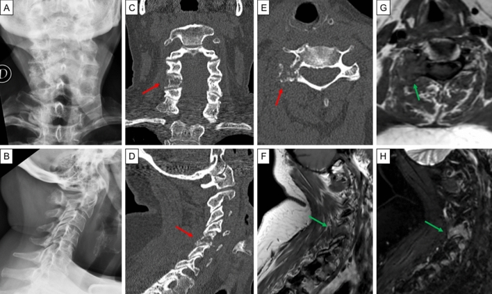
Fig. 2.
Images from a 55-year-old female with poorly differentiated thyroid cancer and cervical pain. The patient underwent cervical spine plain radiograph (a postero-anterior view, b latero-lateral view) that was reported as negative. Therefore, after a few days, she underwent cervical spine CT. The CT multiplanar reconstructions images (c axial view, d coronal view, e sagittal view) showed the presence of an osteolytic bone lesion involving the peduncle and the zygapophyseal joint with minimal involvement of the hemisoma of the fifth cervical vertebra (red arrow). The MR images (f T1-weighted sagittal view; g T1-weighted axial view; h T2-weighted with fat suppression sagittal view) confirmed the CT scan findings, with a lesion which resulted hypointense in T1 and hypertense in T2 (green arrow). CT computed tomography, MR magnetic resonance

