
Figure 4. ZMYND8 inhibits NF-κB activation and expression of IFN-β and ISGs in breast tumors.
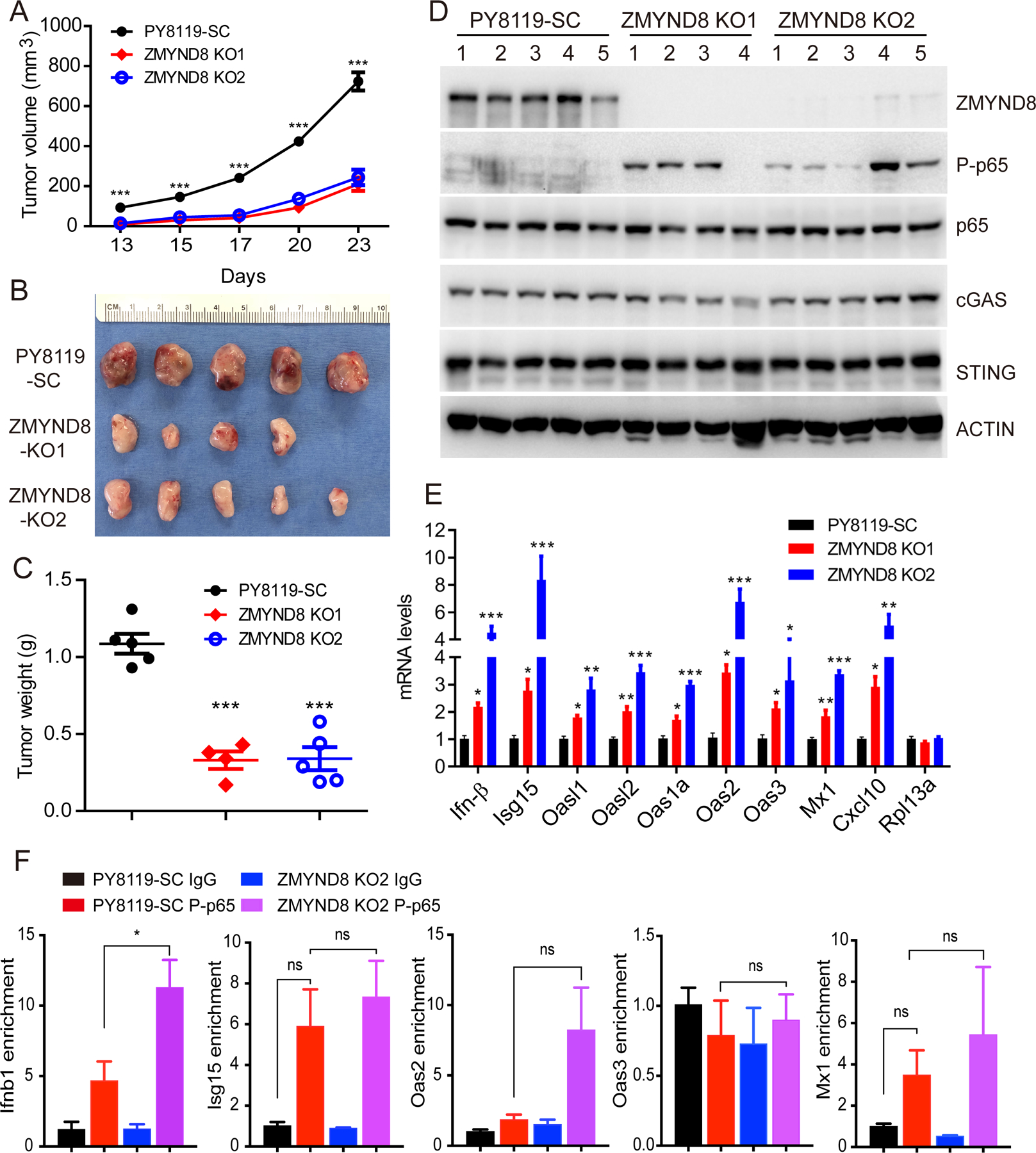
(A-C) SC and ZMYND8 KO PY8119 cells were orthotopically implanted into the mammary fat pad of female C57BL/6J mice, respectively. Tumor growth curve is shown in A (n = 4–5, mean ± SEM). Tumor image on day 23 is shown in B. Tumor weight on day 23 is shown in C (n = 4–5, mean ± SEM). ***p < 0.001 by one-way ANOVA with Dunnett’s test. (D) Immunoblot analysis of indicated proteins in SC and ZMYND8 KO PY8119 tumors. (E) RT-qPCR analysis of IFN-β, ISGs and Rpl13a mRNAs in SC and ZMYND8 KO PY8119 tumors (n = 4–5, mean ± SEM). *p < 0.05; **p < 0.01; ***p < 0.001 vs. SC by one-way ANOVA with Dunnett’s test. (F) ChIP-qPCR analysis of phospho-p65 (P-p65) enrichment at Ifnb1, Isg15, Oas2, Oas3, Mx1 genes in SC and ZMYND8 KO2 PY8119 cells (n = 3, mean ± SEM). *p < 0.05 by two-way ANOVA with Tukey’s test. ns, not significant.