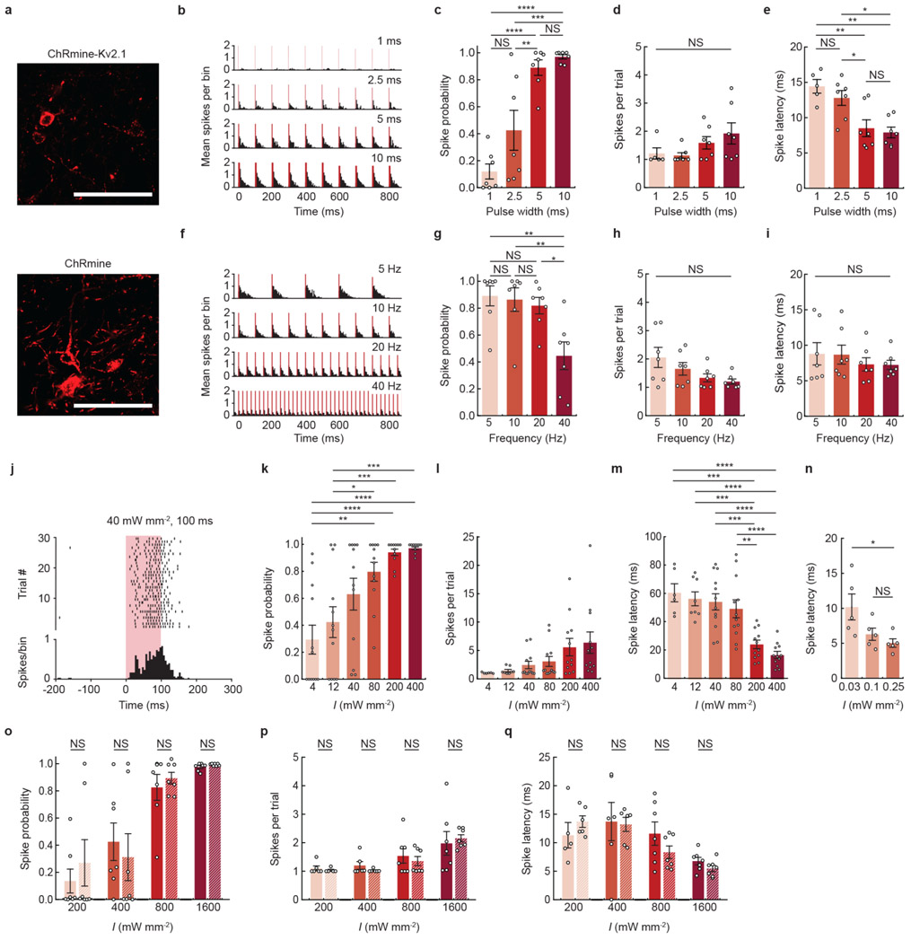
Extended Data Fig. 1 ∣. Extracellular electrophysiological characterization of transcranial deep brain optogenetics at different photostimulation parameters in mice.
a, Confocal image of representative neurons in the VTA expressing either soma-localized (Kv2.1) or non-localized ChRmine-oScarlet. Scale bar: 100 μm. b, Representative peristimulus time histogram of transcranial light-evoked spikes at different pulse widths. Light-responsive single-unit neural recordings at different pulse widths plotted as: probability of one or more evoked spikes (c), number of spikes per pulse (d), and latency to the first spike (e) (n = 7 units from 2 mice; one-way ANOVA with Bonferroni post hoc tests: F(3,24) = 22.51, P = 0.0000004 (c); F(3,22)=2.10, P= 0.13 (d); and F(3,22) = 9.30, P = 0.0004 (e). Note two neurons exhibited no spike response at 1-ms pulse width and therefore do not have an associated latency or spike count at this pulse width). f, Representative peristimulus time histogram of transcranial light-evoked spikes at different frequencies. Light-responsive single-unit neural recordings at different frequencies plotted as: probability of one or more evoked spikes (g), number of spikes per pulse (h), and latency to the first spike (i) (n = 7 from 2 mice; one-way ANOVA with Bonferroni post hoc tests: F(3,24) = 6.31, P = 0.003 (g); F(3,24) = 2.82, P = 0.06 (h); and F(3,24)=0.52, P = 0.67 (i)). j, Representative raster plot and peristimulus time histogram of transcranial light-evoked spikes at low light power (40 mW mm−2, 100 ms duration). Light-responsive single-unit neural recordings at different irradiance (I) with an extended pulse width of 100 ms plotted as: probability of one or more evoked spikes (k), number of spikes per pulse (l), and latency to the first spike (m) (n = 12 units from 2 mice; one-way ANOVA with Bonferroni post hoc tests: (k) F(5,66) = 10.46, P = 0.0000002 (l) F(5,56) = 2.81, P = 0.02, and (m) F(5,56 ) = 14.08, P = 0.000000006. Note six (4 mW mm−2) and four (12 mW mm−2) neurons exhibited no spike response and therefore do not have an associated latency or spike count at these irradiances). n, Latency to first spike determined from patch recordings of cultured hippocampal neurons at low irradiance exhibiting increased time to fire action potentials with decreasing photon density (n = 5 neurons, one-way ANOVA with Bonferroni post hoc tests: F(2,12) = 36, P = 0.03). Light-responsive single-unit neural recordings at different irradiance for ChRmine-expressing neurons with (nonstriped) or without (striped) the Kv2.1 peptide tag:probability of one or more evoked spikes (o), number of spikes per pulse (p), and latency to the first spike (q) (n = 7 units from 2 mice (ChRmine) and n = 7 units from 2 mice (ChRmine-Kv2.1); two-way ANOVA with Bonferroni post hoc tests: ChRmine-Kv2.1 vs ChRmine (o) F(1,48)=0.02, P=0.88, (p) F(1,43)=0.08, P=0.78, and (q) F(1,43)=0.42, P=0.52). No differences in the ability to photoactivate neurons with or without the Kv2.1 tag was observed. In b-e, 635 nm light was delivered at 10 Hz and 800 mW mm−2 light power at different pulse width. In f-i, 635 nm light was delivered at 800 mW mm−2 mW light power with 5-ms pulse width at different frequencies. In j-m, 635 nm light was delivered at 1 Hz with 100 ms pulse at different irradiance. *P < 0.05; **P < 0.01; ***P < 0.001; ****P < 0.0001; NS, not significant. Data are mean ± sem.

