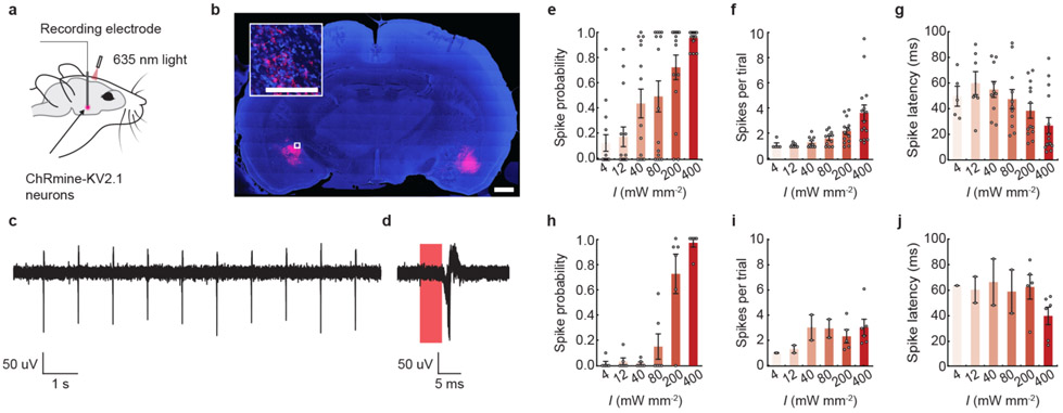
Extended Data Fig. 2 ∣. ChRmine photoactivatable up to 7 mm from the skull.
a, Schematic of experiment for extracellular recording in anesthetized rats. b, Confocal image of a coronal slice from rat depicting DAPI-stained cells (blue) and soma-localized ChRmine-oScarlet-Kv2.1 (red) expression in neurons at 6 mm (left) and 7 mm (right). Inset: expanded view of neurons expressing ChRmine. Scale bar: 1 mm, (inset) 100 μm. c, d, Example voltage trace of light-evoked activity in response to 5-ms pulse width of light delivered at 10 Hz with a 635 nm laser at 800 mW mm-2. Light-responsive single-unit neural recordings at 6 mm plotted as: probability of one or more evoked spikes (e), number of spikes per pulse (f), and latency to the first spike (g) (n = 15 units from 2 rats). Light-responsive single-unit neural recordings at 7 mm plotted as: probability of one or more evoked spikes (h), number of spikes per pulse (i), and latency to the first spike (j) (n = 6 units from 2 rats). In e-g and h-j, 635 nm light was delivered at 1 Hz with a pulse width of 100 ms at different irradiance (I). At 400 mW mm−2, these conditions resulted in a spike probability of 0.96 with an average spike latency of 27 ± 6 ms and a spike count of 3.7 ± 0.6 recorded at 6 mm in depth, and a spike probability of 0.97 with an average spike latency of 39 ± 7 ms and a spike count of 3 ± 0.6 at 7 mm in depth. At the same conditions, no light-evoked activity was recorded at 8 mm deep across 2 rats. Data are mean ± sem.

