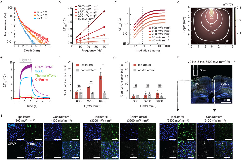
Extended Data Fig. 3 ∣. ChRmine enabled transcranial deep brain optogenetics at light powers that elicit minimal tissue heating.
a, Light transmission profile through brain tissue from a 400-μm 0.39 NA optical fiber calculated from Monte Carlo simulations of photon propagation. b, Calculated maximum temperature change associated with a 635 nm laser delivered at various light powers and frequencies with a pulse width of 5 ms. Note conditions used for transcranial optogenetics in mice (800 mW mm−2 and up to 10% duty cycle) resulted in minimal tissue heating at steady state (~0.3 °C, gray dashed line). c, Calculated maximum temperature change associated with a 635 nm laser delivered at various light powers as a function of continuous irradiation time. Note conditions used for transcranial optogenetics in rats (400 mW mm−2 with 100-ms pulse width at 1 Hz) resulted in minimal tissue heating at steady state (~0.4°C, gray dashed line). d, Modeled temperature distribution in brain tissue at steady state for 635 nm light delivered at 800 mW mm−2, 20 Hz, 5-ms pulse width. e, Calculated maximum temperature change for various laser parameters used for transcranial optogenetics applied for 10 s (see Supplementary Table 2 for summary of parameters). Note stimulation conditions for ChRmine (red) heat tissue less than range reported to have nonspecific temperature effects on behavior (green, 532 nm 222 mW mm−2). Percentage of Iba+ microglia (f) and GFAP+ astrocytes (g) among DAPI-labeled cells within 200 μm of fiber optic source on the photostimulated (ipsilateral, red) or non-photostimulated (contralateral, gray) side. 635 nm light was delivered at 20 Hz with 5 ms pulse at different light powers (800, 3200, and 6400 mW mm−2) for 1 hour. Animals were perfused 24 hours after photostimulation. No glial accumulation was evident at 800 mW mm−2 and 10% duty cycle conditions (n = 3 mice; two-sided paired t-test, *P = 0.03; **P = 0.005; NS, not significant). h, Representative confocal image depicting a coronal slice treated at 6400 mW mm−2 exhibiting tissue lesioning on the ipsilateral side. i, Representative confocal images with indicated side of photostimulation and irradiance. Scale bar: 100 μm. Data are mean ± sem.

