Figure 1.
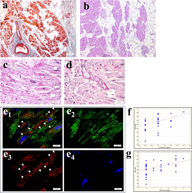
Interstitial area of atrial appendage myocardium of patients with AF. (a,b) Significant fibrosis and lipomatosis in the interstitium of atrial appendage myocardium. (a) Masson’s trichrome stain. (b) Hematoxylin and eosin. bar 7 µm. (c–e) Isolated atrial amyloidosis in atrial appendage myocardium. Amyloid deposition in the interstitial area on the surface of cardiomyocytes (c) and the wall of the intramural vessel (d). (c,d) Sirius red stain. bar 30 µm. e1-4—ANP+ fibrillar material in the interstitium on the surface of cardiomyocytes sarcolemma and in the walls of blood vessels (arrows). ANP+ atrial granules in the sarcoplasm of cardiomyocytes. e1—ANP/Desmin/DAPI immunohistochemistry. e2—Desmin-positive fibrils in the sarcoplasm of cardiomyocytes (Alexa 488). e3—Detection of ANP-positive atrial granules in the sarcoplasm of cardiomyocytes and ANP-positive fibrils in the interstitium (arrows) (Alexa 546). e4—DAPI-stained nuclei. Immunoconfocal laser microscopy. bar 10 µm. (f,g) Correlation of the amyloid distribution in the LAA (f) and RAA (g) myocardium with the age of patients with AF.

