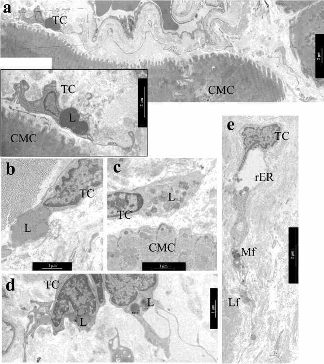
Figure 6.
Lipid inclusions and dystrophic changes in atrial appendage myocardial TCs in patients with AF. (a) Single lipid inclusions in the perinuclear zone of the TC cytoplasm. bar 1 µm. Insert—bar 2 µm. (b–d) The accumulation of multiple small and large lipid inclusions in the perinuclear zone of the TCs cytoplasm. bar 1 µm. (e) Myelin-like figures (Mf), rough endoplasmic reticulum (rER) and lipofuscin granules (Lf) in the TC cytoplasm. bar 2 µm. TEM. TC—telocyte, CMC—cardiomyocyte. L—lipid inclusions.

