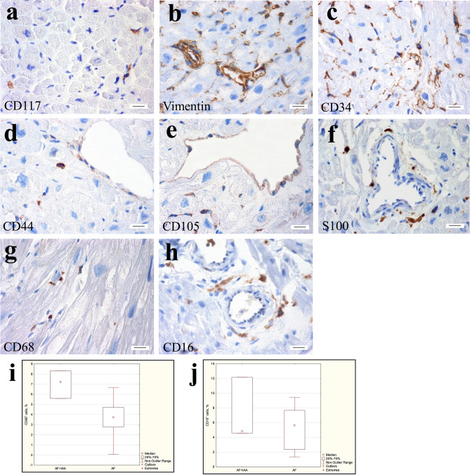
Figure 7.
Immunohistochemical detection of TCs in atrial appendage myocardium of patients with AF. (a) Single CD117+ cells in the interstitium. (b) Vimentin+ TCs, endotheliocytes, and connective tissue cells. (c) CD34+ endotheliocytes, and TCs surrounding vessels, cardiomyocytes, and endotheliocytes in the interstitium. (d) CD44+ TCs in the interstitium, as well as weakly-labeled endotheliocytes. (e) CD105+ endotheliocytes. (f) S100-positively stained nerve fibers. (g) CD68+ round cells of histiocytic origin and spindle-shaped TCs in the interstitium. (h) CD16+ round cells of histiocytic origin and spindle-shaped TCs in the perivascular area. (a–h) Streptavidin–biotin complexes. bar 15 µm. (i) The number of CD68+ cells in the atrial appendage myocardium of the AF + IAA patients significantly exceeds the number of these cells in the AF group. Mann–Whitney test, p < 0.05. (j) The number of CD16+cells in the atrial appendage myocardium of the AF + IAA patients does not differ significantly from the number of these cells in the AF group. Mann–Whitney test, p > 0.05.

