FIGURE 7.

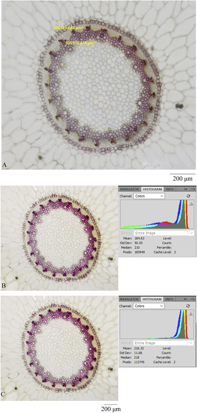
Transverse section was processed by different microscopes and software: (A) ZEISS and ZEN, (B) OLYMPUS and photoshop (showing the pixel of xylem and pith), and (C) OLYMPUS and photoshop (showing the pixel of pith).

Transverse section was processed by different microscopes and software: (A) ZEISS and ZEN, (B) OLYMPUS and photoshop (showing the pixel of xylem and pith), and (C) OLYMPUS and photoshop (showing the pixel of pith).