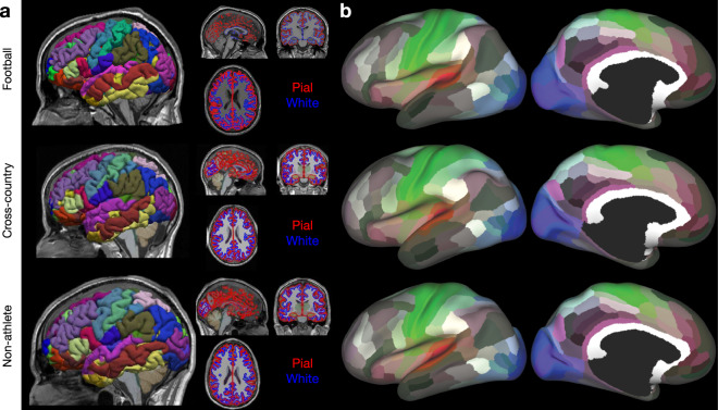
Fig. 2.
Anatomical (T1w) preprocessing: Freesurfer and 180 node multimodal atlas mapping (a) Representative images from each group of the Freesurfer outputs: pial (red) and white (blue) matter surfaces, and the aparc.a2009s + aseg (i.e. Destrieux) parcellation. Images were generated using brainlife.io’s Freeview viewer. (b) Representative images from each group of the 180-node multimodal (hcp-mmp) atlas mapped to an inflated representation of the cortical surface. Images were generated using brainlife.io’s Connectome Workbench viewer.

