Figure 2.
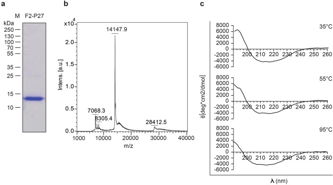
Characterization of recombinant F2-P27 protein. (a) Coomassie brilliant blue-stained SDS-PAGE showing purified F2-P27 subunit. Molecular weights (kDa) are indicated on the left margin. (b) Mass spectrometry of recombinant F2-P27 subunit. The mass/charge (m/z) ratios are shown on the x-axes, and the intensities are displayed on the y-axes in arbitrary units (a.u.). (c) Far-UV CD spectra of F2-P27 subunit at different temperatures (35°C, 55°C and 95°C). Mean residue ellipticities (θ) (y-axes) at given wavelengths (x-axes).

