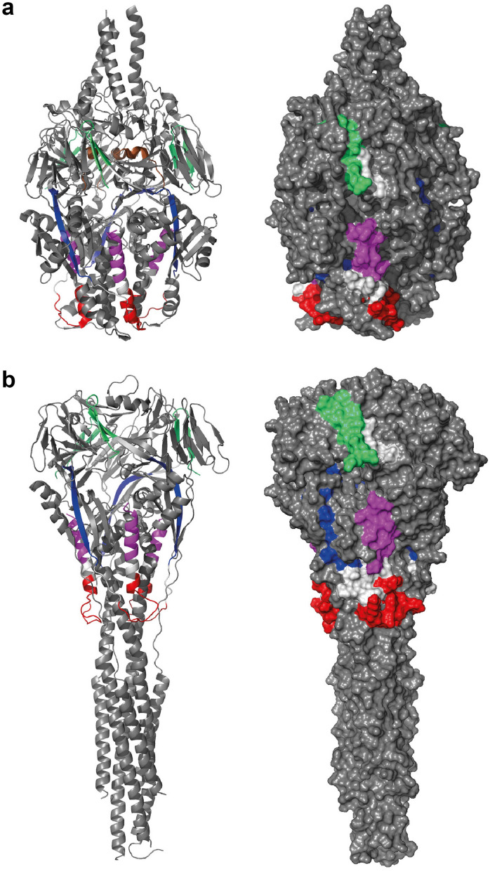
Figure 7.
The surface exposure of the peptides in the folded forms of the RSV-F protein. (a) the pre-fusion F protein (PDB 3RRR) and (b) the post-fusion F protein (PDB 6APD) are shown in a cartoon-presentation (left panel) and as a surface presentation (right panel). Both forms exist as trimers with the three-fold axis aligned with the vertical axis. The peptides have been colored (P1 green, P2 blue, P3 red, P4 violet and P6 brown; the atomic coordinates for P5 and P6 are missing completely in the post-fusion F protein structure and only 10 residues of P6 have been determined in the pre-fusion F protein structure (brown α-helix in a). P1, P3 and P4 display several surface exposed regions, whereas P2 is mainly buried.

