Figure 2.
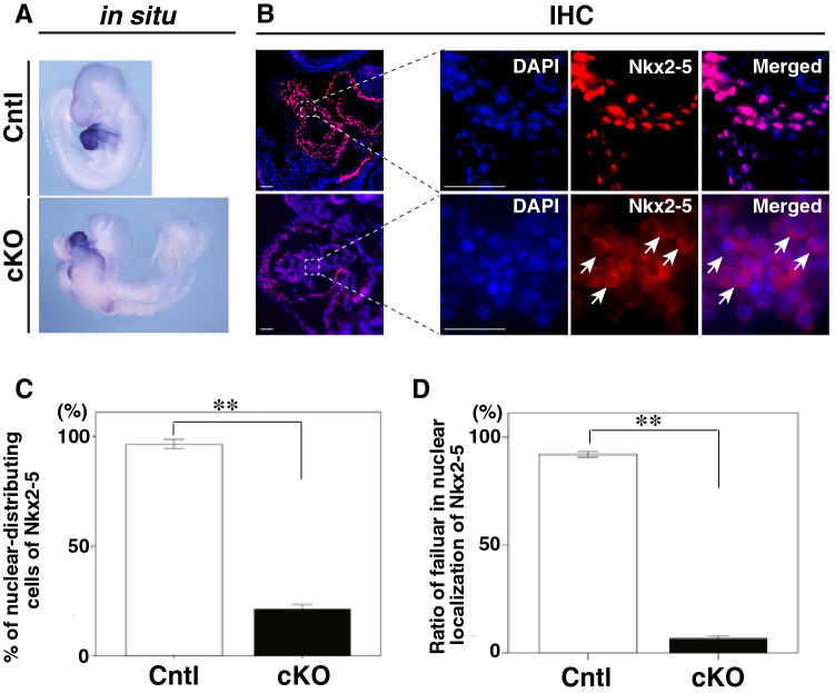
Lack of nuclear localization of Nkx2-5 in Smad4-cKO embryonic hearts. (A) Whole-mount ISH of control and Smad4-cKO embryos with a probe for Nkx2-5 at E9.5. (B) Immunofluorescent staining of sagittal sections of control and Smad4-cKO embryos at E9.5 using anti-Nkx2-5 antibodies (red) and DAPI for detection of nuclei (blue). Arrows indicate the lack of nuclear localization of Nkx2-5. H, heart. Scale bar = 100 μm. (C and D) Percentage of Nkx2-5 protein distributing cell in control and Smad4-cKO embryonic hearts (C) and the percentage of cells showing nuclear localization of Nkx2-5 in control and Smad4-cKO embryonic hearts (D). Data were collected from 10 sections for each embryo (n = 3 and 3 for control and Smad4-cKO, respectively). Data are presented as mean ± SEM, **P < 0.01 (Mann–Whitney U test).

