Figure 4.
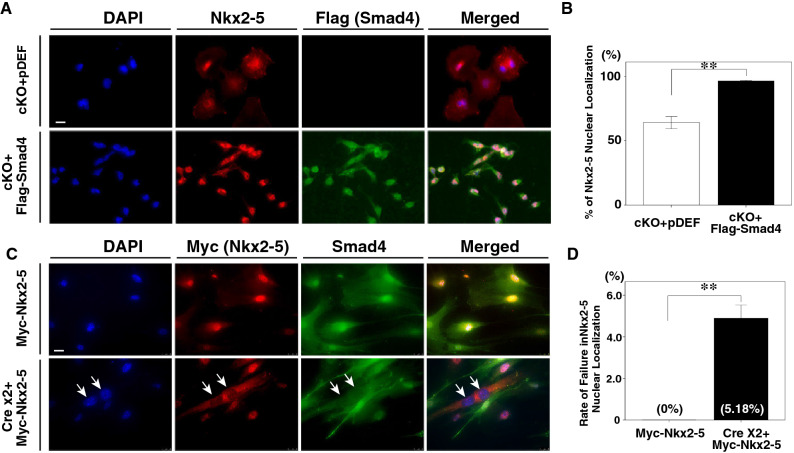
Introduction of Smad4 rescues the nuclear localization of Nkx2-5 in mutant cells and depletion of the Smad4 gene blocks the nuclear translocation of Nkx2-5 in fibroblast cells. (A) Double immunohistochemistry with anti-Nkx2-5 (red) and anti-Flag (green) antibodies. Cells from Smad4-cKO embryonic hearts were transfected with an empty vector (pDEF) as a control (upper panels) or Flag-tagged Smad4 expression vector (lower panels). Arrows show the failure of Nkx2-5 to localize to the nucleus. DAPI was used to stain nuclei. (B) Percentage of cells showing nuclear localization of Nkx2-5. Nuclear localization of Nkx2-5 was significantly increased in the presence of Smad4, compared to cells transfected with empty vector. Data were collected from 20 slides for each experiment (n = 4 and 4 for control and Smad4-cKO, respectively). Data are presented as mean ± SEM. *P < 0.01 (Mann–Whitney U test). (C) Double immunohistochemistry with anti-Myc (red) and anti-Smad4 (green) antibodies. Fibroblast cells from Smad4f./f embryos were transfected with the Myc-Nkx2-5 expression vector and showed nuclear localization of Nkx2-5. After depleting Smad4 by transfecting fibroblasts with Cre expression vector twice, Nkx2-5 was no longer localized to the nucleus. DAPI was used to stain nuclei. (D) The proportion of non-nuclear localization of Nkx2-5 in control (transfected with Myc-Nkx2-5) and Smad4-cKO (transfected with Myc-Nkx2-5 after two rounds of Cre expression vector transfection) cells. Data were collected from 20 slides for each experiment (n = 3 and 3 for control and Smad4-cKO, respectively). Data are presented as mean ± SEM. **P < 0.01 (Mann–Whitney U test).

