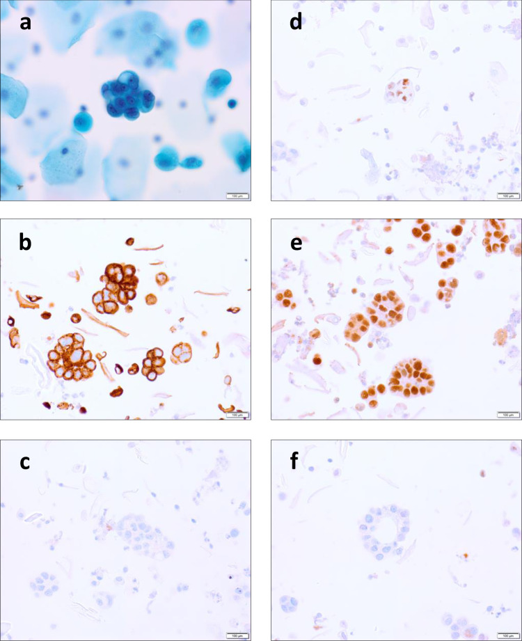
Fig. 2. Photomicrograph showing morphology and immunohistochemistry profile of endometrial cancer cells in urogenital samples.
a Urine sample showing positive urine cytology (SurePath), demonstrating a single cluster of endometrial cells with malignant features, including enlarged hyperchromatic nuclei, coarse chromatin, prominent nucleoli, irregular nuclear outlines and high nuclear-to-cytoplasmic ratio. b–f Vaginal sample demonstrating the immunoprofile of these cells, consistent with epithelial cells from the female genital tract, CK7+ (b), CK20− (c), ER+ (d), PAX8+ (e), GATA3− (f). Scale bar = 100 μm.

