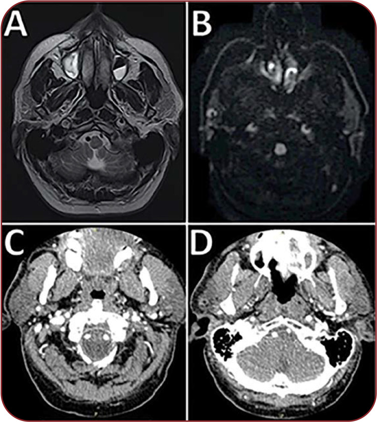
FIGURE 2.
(A) Cervical magnetic resonance imaging scan (cross section) T2-weighted hypersignal present in the walls of C1-C5 segments of the right internal carotid artery and of C1-C2 segments of the left internal carotid artery (suggestive for intramural hematomas), and dissection fold that occludes the lumen of the right carotid artery. (B) Cervical magnetic resonance imaging scan (cross section) – diffusion-weighted imaging – revealing the semilunar shape of the bilateral internal carotid artery intramural hematomas. (C and D) Computed tomography angiography of the supra-aortic trunks showing patent lumina of both internal carotid arteries.

