Fig. 1.
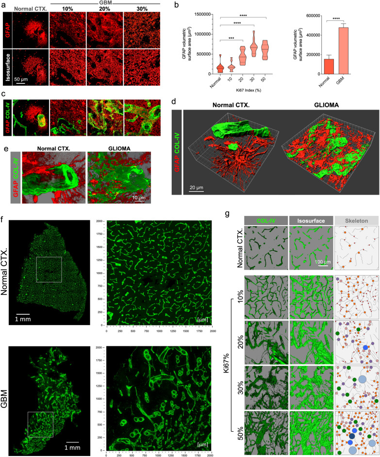
Vascular map and GFAP’s altered spatial relationship in GBM. a GFAP distribution of characteristic protoplasmic astrocytes in normal CTX, and glioma cells in GBM-affected cases, displayed on original confocal images (top panel) and the corresponding rendered isosurfaces (bottom panel). b GFAP-expressing cells show a massive increase in GBM as reflected in volumetric surface area analysis relative to Ki67% and all tumor cases grouped. c Visualization of correlative vasculature associated with GFAP+ cells in normal brain and GBM. Merged views of GFAP from panel A, and blood vessels, evidenced by COL-IV (green) are shown. d 3D rendering of GFAP and COL-IV in normal CTX and GBM highlight the differences between normal and GBM blood vessels in relation with surrounding GFAP-expressing cells. e Detailed, higher magnification 3D renderings show altered spatial relationship of basement membrane and GFAP. f Hypermosaic of cortical sample of a normal brain CTX and GBM, and high-resolution mosaics from white inset. g Confocal alpha blending, 3D rendering of high-magnification images and skeletonization (including branching points color-coded by size) reveal dramatic changes in blood vessel distribution and architecture relative to increasing Ki67 index in GBM

