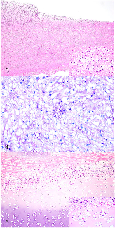
Figures 3-4.
Mucopolysaccharidosis type VI, aorta, Great Dane puppy. Figure 3. The wall of the aorta is thickened and the intima is expanded by proliferating fibroblasts forming plaque-like lesion. Inset. The smooth muscle myocytes within the wall of the aorta contain cytoplasmic vacuoles. Hematoxylin and eosin. Figure 4. The vacuoles within the smooth muscle myocytes are mostly clear but occasionally stain metachromatically, consistent with accumulation of glycosaminoglycan. Toluidine blue.
Figure 5. Mucopolysaccharidosis type VI, tracheal cartilage, Great Dane puppy. The tracheal cartilage is thickened with irregular chondrones and surrounded by proliferating fibroblasts. Inset. The chondrocytes and fibroblasts within the wall of the trachea contain cytoplasmic vacuoles. Hematoxylin and eosin.

