Fig. 6.
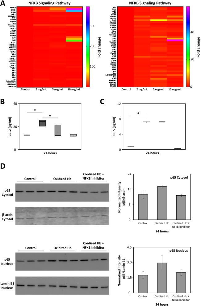
Oxidized Hb induces NFKB signaling pathway. Mixed glial cells were exposed to increasing concentration of oxidized Hb (2–10 mg/ml) or culture medium only (control) for 24 h. a A heat map revealing a concentration-dependent upregulation by oxidized Hb in mRNA expression of activators, transcriptional factors, and pro-inflammatory cytokines associated with the NFKB pathway. Addition of the NFKB activation inhibitor VI benzoxathiole (3.0 μM) in the presence of oxidized Hb (2.5 mg/ml, grey bar, n = 5) partly inhibited the oxidized Hb (dark grey bar, n = 5) induced protein level of CCL2 (b), but not CCL5 (c). Results of b and c are presented as box plots displaying medians and 25th and 75th percentiles. Control (white bar, n = 5) and NFKB activation inhibitor VI benzoxathiole only (light grey, n = 5). Protein levels of the NFKB transcription factor p65 were assessed in the cytosolic and nuclear fraction using western blot. A tendency towards increased level of p65, in both the cytosolic and the nuclear fraction, was observed following exposure to oxidized Hb (2.5 mg/ml). Co-administration of the NFKB inhibitor VI benzoxathiole displayed a trend of reduced p65 protein levels (d). β-actin (cytosolic fraction) and Lamin B1 (nuclear fraction) were used as protein loading controls and analyzed on the same material as p65. All samples were applied to one membrane. Membranes are from a representative analysis. Densitometric quantification was performed and mean normalized intensity, normalized for respective β-actin (cytosolic) or Lamin B1 (nuclear) intensity, is presented. Differences between oxidized Hb vs. control, oxidized Hb + NFKB inhibitor vs. control, and oxidized Hb + NFKB inhibitor vs. oxidized Hb were analyzed using the Kruskal–Wallis test followed by pairwise comparison with significance values adjusted for multiple comparisons. *P < 0.05

