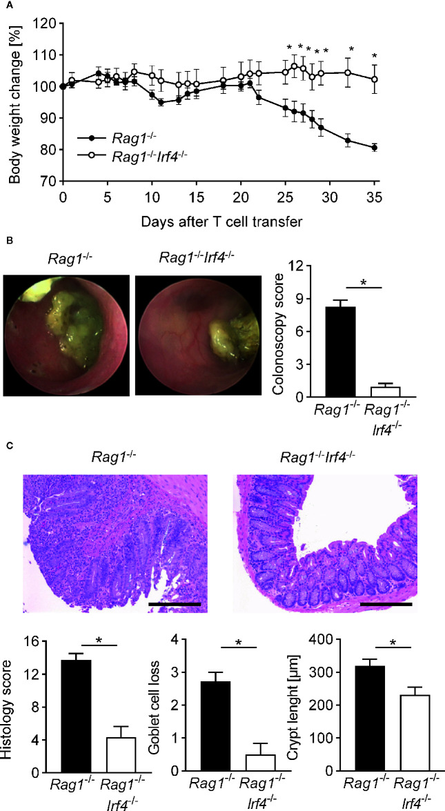
Figure 1.
Inactivation of IRF4 in non-T cells abrogates clinical, endoscopic, and histopathological signs of colitis. At day 0 Rag1 −/− and Rag1 − / − Irf4 −/− mice were injected i.p. with 1 × 10⁶ naïve (CD4+CD25−) T cells. (A) Percentage body weight changes compared to the original body weight at day 0 were assessed over time. A representative course of body weight changes per experimental group from one of four experiments is shown (Rag1 −/− n = 6; Rag1 − / − Irf4 −/− n = 5). (B) Colitis severity was analyzed by colonoscopy when T cell recipient Rag1 − / − mice showed sustained weight loss of more than 10% of their initial body weight for at least one week (four to five weeks after T cell transfer). Colonoscopy score of three independent experiments and representative endoscopic images for every experimental group are shown. Rag1 −/− and Rag1 − / − Irf4 −/− mice were sacrificed when colitis was established in T cell recipient Rag1 −/− mice identified by weight loss and colonic inflammation via colonoscopy (five to six weeks after T cell transfer). (C) Histopathological scoring of the inflammation present in the distal colon. One representative H&E-stained histopathological cross-section of the distal colon per group is shown. Scale bars: 100 µm. (B, C) Data are combined from three individual experiments (Rag1 −/− n = 11; Rag1 − / − Irf4 −/− n = 12). Data were analyzed by Student’s t test and are shown as mean ± SEM. *p <0.05.

