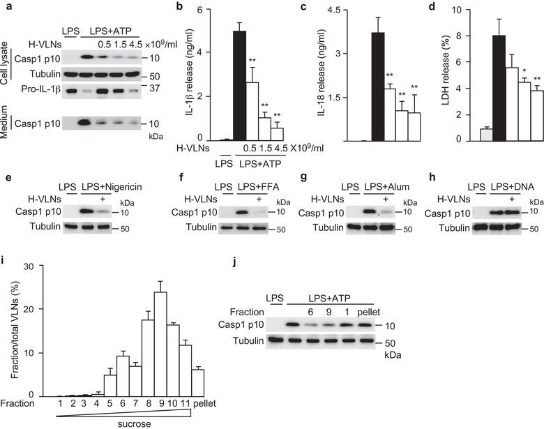
FIGURE 2.

H‐VLNs specifically suppressed activation of the NLRP3 inflammasome. [a‐d. The primary macrophages were pretreated with H‐VLNs for 16 h, followed by addition of LPS + ATP to activate the NLRP3 inflammasome. H‐VLNs inhibited the level of Casp1 p10 in the cell lysate and culture medium (a), IL‐1β secretion (b), IL‐18 secretion (c), and pyroptosis (d). Lactate dehydrogenase (LDH) release assay was used to indicate the level of pyroptosis. e‐h. H‐VLNs inhibited Casp1 autocleavage when the NLRP3 inflammasome was activated with nigericin (e), free fatty acid (FFA) (f), or alum (g). H‐VLNs had marginal effects on Casp1 autocleavage upon AIM2 inflammasome activation (h). In e‐h, 4.5×10⁹/ml of H‐VLNs were used. i‐j. H‐VLNs were fractionated using sucrose gradient. Fractions were collected at 1 ml each and named as fractions 1‐11 from top to bottom. The particle number in each fraction was measured and presented as a percentage of the total particle numbers (i). The particles in fractions 6 and 9, the light particles in fraction 1, and the aggregates in pellet were collected and assessed for their inhibitory effects on NLRP3 inflammasome‐mediated Casp1 autocleavage (j). Data were presented as mean ± SEM. N = 3. *P < 0.05, **P < 0.01 relative to BMDMs treated with LPS + ATP (black bar). Tubulin showed equivalent loading of cell lysates].
